The keys to a major process in DNA repair
Researchers from the Institut Jacques Monod (CNRS/University of Paris Diderot), the Institute of Biology of the Ecole Normale Supérieure (ENS/CNRS/Inserm), and the University of Bristol, have described for the first time in its totality the mechanisms by which DNA damaged by UV radiation is repaired, and how the proteins involved in this process cooperate to ensure its efficiency. This work opens new perspectives not only in the fight against cancer but also in combating certain bacterial infections, and is published in Nature on August 3rd 2016.1
- 1See the two previous articles: Initiation of transcription-coupled repair characterized at single-molecule resolution, Kevin Howan et al., Nature, 2012 and A dynamic DNA-repair complex observed by correlative single-molecule nanomanipulation and fluorescence, Evan T. Graves et al., Nature Structural and Molecular Biology, 2015
The DNA of our cells is continuously damaged by numerous external agents, such as carcinogens contained in tobacco smoke or UV radiation emitted by the sun. If left unrepaired, this damage leads to mutations which ultimately favor the emergence of cancerous cells, which is why the cell must rapidly and efficiently repair its DNA. To do so, the cell employs a battery of enzymes which must act in a synchronous fashion to identify and repair the damaged parts of its genome. The complexity of this process has long stumped researchers trying to understand the mechanisms at play.
Thanks to new nanotechnologies, a team of scientists which brings together both physicists and biologists has been able to film, in real-time, the enzymes that repair DNA damage. This work started in 2012, when the team focused on the initial steps of the DNA repair mechanism. Today the team has revealed, for the first time, the repair process in its entirety.
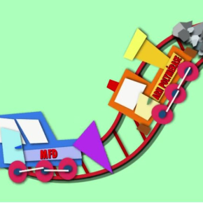
A special type of microscope, which makes it possible to both manipulate and observe single molecules of DNA and proteins, has enabled the team to observe a single DNA molecule, damaged by UV. They added to it the RNA polymerase enzyme, the one naturally responsible for “reading” the lengths of the DNA code and initiating production of protein from this DNA code, but which can get “stalled” if it reads a segment of damaged DNA. It is thanks to this “stalling” that the cell recognizes that the DNA has been damaged and launches its repair. In practical terms, the team of scientists was able to observe a series of four proteins (named Mfd, UvrA, UvrB and UvrC) successively interacting with the RNA polymerase and coordinating among themselves and the UV-damaged DNA to enact the latter's repair.
By determining the order in which these components acted and by characterizing the manner in which they “handed off” to each other in a kind of molecular relay-race, the team was able to define the critical steps of this process.
This work will ultimately lead to new applications, both in the fight against cancer and in efforts to treat pathogenic bacteria. Indeed, when cancer cells become resistant to chemotherapy or radiation therapy – the purpose of which is to damage the DNA of cancer cells – it is because these cancer cells have activated DNA repair and undone clinically-generated DNA damage. One can thus work towards preventing DNA repair during cancer therapy so as to prevent tumor resistance to therapy. It also turns out that some pathogenic bacteria, including those responsible for tuberculosis, use proteins very similar to Mfd to proliferate. Thus, identifying how these proteins work together to enact DNA repair could also be useful in fighting pathogenic bacteria.
To learn more about this process, watch the video (in French): Proteins which repair DNA, produced by CNRS Images (2012, 7 minutes).
Reconstruction of bacterial transcription-coupled repair at single-molecule resolution, Jun Fan, Mathieu Leroux-Coyau, Nigel J. Savery, Terence R. Strick. Nature, 3 August 2016. View web site


